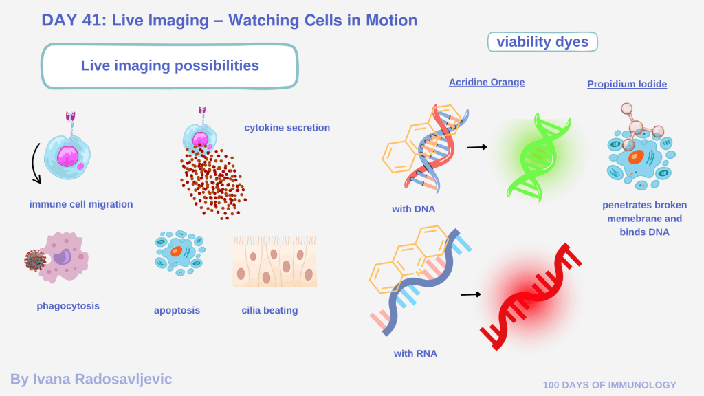
Live-cell imaging is one of the most captivating technique in modern cell biology and immunology. Unlike fixed imaging, which captures a single frozen moment, live imaging allows us to observe the dynamic behavior of cells such as migration, phagocytosis, cytokine secretion, apoptosis, or cilia beating 𝘪𝘯 𝘳𝘦𝘢𝘭 𝘵𝘪𝘮𝘦 [1].
𝗧𝗵𝗲 𝗥𝗼𝗹𝗲 𝗼𝗳 𝗩𝗶𝗮𝗯𝗶𝗹𝗶𝘁𝘆 𝗗𝘆𝗲𝘀 𝗶𝗻 𝗟𝗶𝘃𝗲 𝗜𝗺𝗮𝗴𝗶𝗻𝗴
In live-cell studies, viability dyes distinguish between living and dead cells. Common examples include 𝘈𝘤𝘳𝘪𝘥𝘪𝘯𝘦 𝘖𝘳𝘢𝘯𝘨𝘦 (𝘈𝘖) and 𝘗𝘳𝘰𝘱𝘪𝘥𝘪𝘶𝘮 𝘐𝘰𝘥𝘪𝘥𝘦 (𝘗𝘐).
Acridine Orange is a nucleic acid-selective fluorescent dye that permeates both live and dead cells. It intercalates into double-stranded DNA and binds electrostatically to single-stranded RNA, emitting green fluorescence for DNA and red for RNA. Allows visualization of overall viability [2].
Propidium Iodide, cannot penetrate intact membranes. It fluoresces red only when bound to DNA in cells with compromised membranes, thus identifying dead or late-apoptotic cells [3].
In flow cytometry, AO and PI can interfere with downstream analyses, such as RNA extraction or live-cell recovery, due to their intercalating and phototoxic properties. For viable cell sorting, more stable alternatives like Calcein AM or SYTO dyes are preferred.
𝗔𝗻𝗲𝗰𝗱𝗼𝘁𝗲 𝗳𝗿𝗼𝗺 𝘁𝗵𝗲 𝗟𝗮𝗯
Just before Christmas 2023: I was working with a Master’s student on differentiating Normal Human Bronchial Epithelial (NHBE) cells. After weeks of culturing, we decided to take a look under the brightfield microscope. Suddenly, we saw the cilia were moving! The fact that they moved meant our basal cells had fully differentiated – a huge success. Excited, we called our supervisor, who joined us in the lab celebration. He brought a live-cell viability dye to confirm that what we saw was indeed living, beating cilia.
𝗟𝗶𝘃𝗲 𝗜𝗺𝗮𝗴𝗶𝗻𝗴 𝗶𝗻 𝗜𝗺𝗺𝘂𝗻𝗼𝗹𝗼𝗴𝘆 𝗮𝗻𝗱 𝗜𝗺𝗺𝘂𝗻𝗼𝘁𝗵𝗲𝗿𝗮𝗽𝘆
In immunology, live imaging is essential for studying:
Immune cell migration and chemotaxis in inflamed tissue models.
Real-time cytotoxicity assays, where CAR-T or NK cells engage tumor cells.
Macrophage phagocytosis and antigen presentation dynamics.
Calcium flux imaging in T-cell activation studies [4].
𝗤𝘂𝗲𝘀𝘁𝗶𝗼𝗻 𝗳𝗼𝗿 𝘁𝗵𝗲 𝗔𝘂𝗱𝗶𝗲𝗻𝗰𝗲 What is the most fascinating live-cell imaging experiment you’ve ever seen?
Stay tuned for 𝗗𝗮𝘆 𝟰𝟮: 𝗨𝗻𝗹𝗼𝗰𝗸𝗶𝗻𝗴 𝘁𝗵𝗲 𝗣𝗼𝘄𝗲𝗿 𝗼𝗳 𝗠𝘂𝗹𝘁𝗶𝗽𝗹𝗲𝘅𝗶𝗻𝗴 𝗙𝗿𝗼𝗺 𝗖𝘆𝘁𝗼𝗸𝗶𝗻𝗲 𝗣𝗿𝗼𝗳𝗶𝗹𝗶𝗻𝗴 𝘁𝗼 𝗖𝗔𝗥-𝗧 𝗣𝗼𝘁𝗲𝗻𝗰𝘆
𝗥𝗲𝗳𝗲𝗿𝗲𝗻𝗰𝗲𝘀
1. DOI: 10.1126/science.1082160
2. https://www.researchgate.net/publication/313052178
3. DOI: 10.1038/nprot.2006.238
4. doi: 10.1007/s10875-010-9393-6
#100DaysOfImmunology #LiveCellImaging #Microscopy #CellBiology #Immunotherapy #FlowCytometry #CARResearch #CellViability #AcridineOrange #PropidiumIodide
