Fluorescence microscopy is one of the most powerful techniques in modern immunology, allowing visualization of subcellular structures, protein localization, and real-time processes such as cell signaling, apoptosis, or immune cell activation [1].
𝗧𝘆𝗽𝗲𝘀 𝗼𝗳 𝗙𝗹𝘂𝗼𝗿𝗲𝘀𝗰𝗲𝗻𝗰𝗲 𝗠𝗶𝗰𝗿𝗼𝘀𝗰𝗼𝗽𝘆
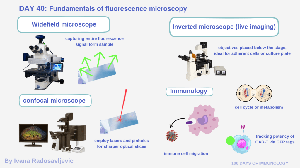
The main types include 𝘸𝘪𝘥𝘦𝘧𝘪𝘦𝘭𝘥 𝘧𝘭𝘶𝘰𝘳𝘦𝘴𝘤𝘦𝘯𝘤𝘦, 𝘤𝘰𝘯𝘧𝘰𝘤𝘢𝘭, 𝘵𝘸𝘰-𝘱𝘩𝘰𝘵𝘰𝘯, 𝘴𝘶𝘱𝘦𝘳-𝘳𝘦𝘴𝘰𝘭𝘶𝘵𝘪𝘰𝘯 (𝘚𝘛𝘌𝘋, 𝘚𝘐𝘔, 𝘗𝘈𝘓𝘔), 𝘢𝘯𝘥 𝘪𝘯𝘷𝘦𝘳𝘵𝘦𝘥 𝘧𝘭𝘶𝘰𝘳𝘦𝘴𝘤𝘦𝘯𝘤𝘦 𝘮𝘪𝘤𝘳𝘰𝘴𝘤𝘰𝘱𝘺, each differing in resolution and optical sectioning [2]. Widefield microscopes capture the entire fluorescence signal from the sample simultaneously, while confocal microscopes employ lasers and pinholes for sharper optical slices. Inverted fluorescence microscopes, often used for live-cell imaging, have their objectives placed below the stage, making them ideal for adherent cells or culture plates [3].
𝗔𝗻𝗲𝗰𝗱𝗼𝘁𝗲 𝗳𝗿𝗼𝗺 𝗠𝘆 𝗝𝗼𝘂𝗿𝗻𝗲𝘆
I was lucky to work with a Zeiss Axio Imager Widefield Fluorescence Microscope valued at over €40,000 at Universitätsklinikum Göttingen. Alone in a dark room, I captured mesmerizing images of cardiomyocytes undergoing apoptosis following mRNA methylation experiments.
Later, during my time at University of Ulm, I used an even more exceptional system – a custom-built inverted fluorescence microscope, assembled from discontinued components. I spent countless hours – sometimes entire Saturdays – counting individual mucus-secreting goblet cells on transwell filters. Each filter contained thousands of cells, and each fluorescent dot represented weeks of culture and care. Though tedious, if I noticed proper vesicles or cilia, those moments were among the most rewarding moments of my scientific path.
𝗦𝗰𝗶𝗲𝗻𝘁𝗶𝗳𝗶𝗰 𝗥𝗲𝗹𝗲𝘃𝗮𝗻𝗰𝗲
Microscopy is indispensable in immunology – it allows visualization of immune cell migration, receptor internalization, cytokine release, or antigen uptake. Fluorescent co-localization studies can confirm whether two proteins interact or share subcellular compartments [4]. In CAR-T and immunotherapy research, microscopy supports potency assays, cell viability checks, and transduction validation by visualizing expression of fluorescent markers such as GFP-tagged chimeric receptors [5].
𝗤𝘂𝗲𝘀𝘁𝗶𝗼𝗻 𝗳𝗼𝗿 𝘁𝗵𝗲 𝗔𝘂𝗱𝗶𝗲𝗻𝗰𝗲: Do you have any anecdotes from the dark room?
Stay tuned for 𝗗𝗮𝘆 𝟰𝟭: 𝗟𝗶𝘃𝗲 𝗜𝗺𝗮𝗴𝗶𝗻𝗴 – 𝗪𝗮𝘁𝗰𝗵𝗶𝗻𝗴 𝗖𝗲𝗹𝗹𝘀 𝗶𝗻 𝗠𝗼𝘁𝗶𝗼𝗻
𝗥𝗲𝗳𝗲𝗿𝗲𝗻𝗰𝗲𝘀
1. DOI: 10.1038/nmeth817
2. http://dx.doi.org/10.1007/978-0-387-45524-2
3. https://doi.org/10.1002/0471732877.emd291
4. DOI: 10.1038/nrm2748
5. DOI: 10.1038/s41568-020-00323-z
#FluorescenceMicroscopy #Immunology #CellBiology #MicroscopyMagic #CARresearch #ScientificImaging #100DaysofImmunology #CellCulture #LiveCellImaging #ZeissMicroscopy
